


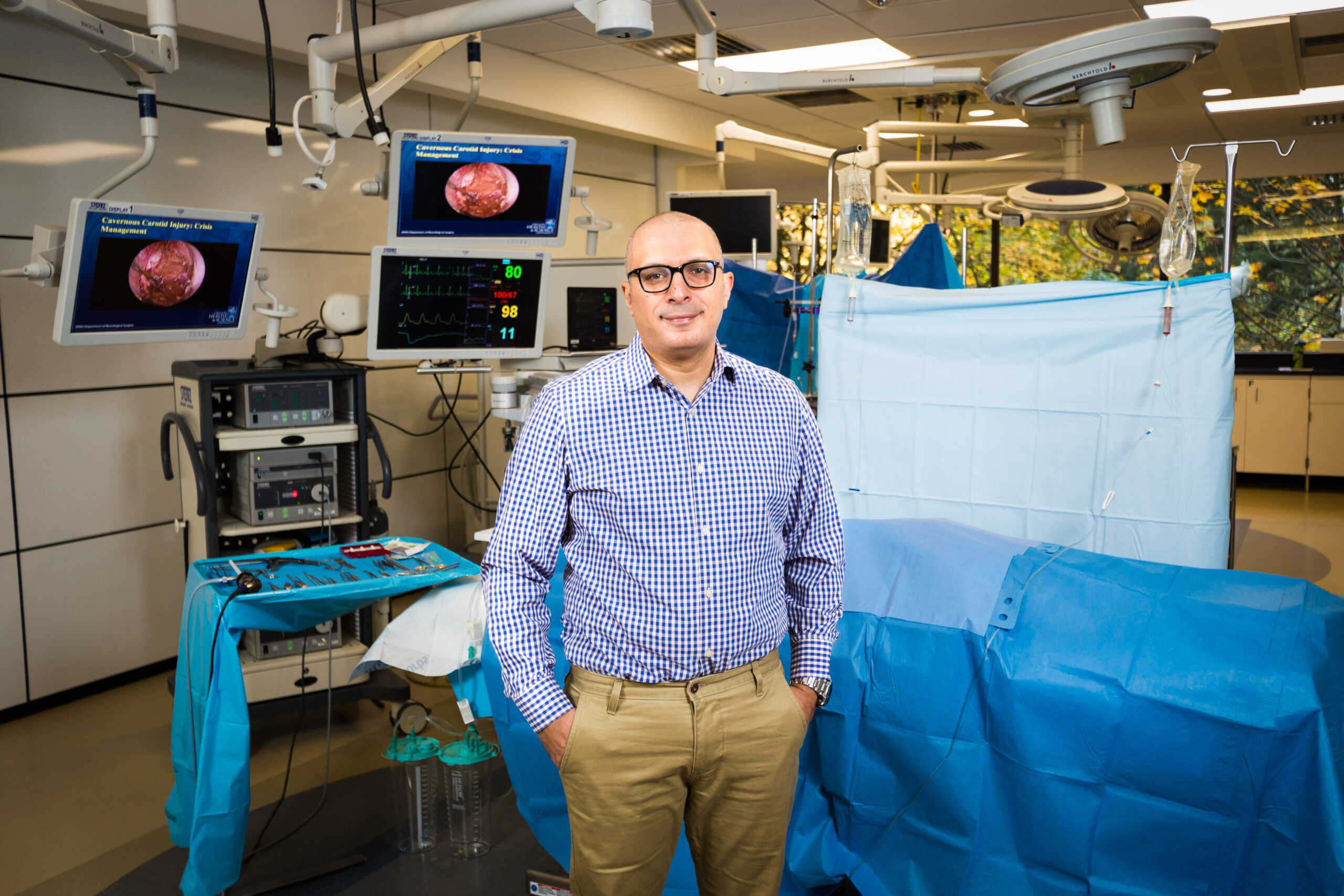
OHSU’s Ahmed Raslan, M.D., is part of a team that has created a new way to look inside the brain during surgery, a breakthrough that is transforming neurosurgery — and our understanding of the brain. The team recently demonstrated that a dense array of sensors can record electrical signals directly from the surface of the brain in astonishing detail. The new brain sensors feature densely packed grids of either 1,024 or 2,048 embedded electrocorticography (ECoG) sensors. The paper describing their breakthrough was published by the journal Science Translational Medicine in January, 2022.
Brain surgery, 100x clearer
The thin, pliable grids of ECoG sensors offer neurosurgeons information — potentially in real-time — on the brain’s cortex in 100 times higher resolution than what was previously possible. Being able to record brain signals at such high resolution allows neurosurgeons to remove as much of a brain tumor as possible while minimizing damage to healthy brain tissue. In the case of epilepsy, higher resolution brain-signal recording capacity could improve a neurosurgeon’s ability to precisely identify the regions of the brain where the epileptic seizures are originating, so that these regions can be removed without touching tissue not involved in seizure initiation.
“The technology will be very helpful in patients with epilepsy and brain tumors, but that’s only the beginning,” said Raslan. “It has the potential to help patients with Parkinson’s disease, Alzheimer’s disease, stroke – the list goes on.”
Support advances in brain health.

Moving with the brain
The human brain is always moving. With each heartbeat, for example, the brain moves with the pulsating blood flowing through it. The platinum nano-rod based sensor grids are thinner and more flexible than today’s clinically approved ECoG grids. The thinness and flexibility allows the sensor grids to move with the brain, enabling a closer connection and better readings. In addition, the grids are manufactured with small, ring-shaped holes that allow cerebral spinal fluid to pass through. In this way, these perfusion holes support a better interface between the sensor grid and the brain surface by allowing the sensor to easily and safely displace the fluid.
The new platinum nano-rod brain sensor grids are ten micrometers thick, approximately one tenth the size of a human hair and 100 times thinner than the one millimeter thick and clinically approved ECoG grids. The nano-rods are embedded in a transparent, soft and flexible biocompatible material called parylene which is in direct contact with the surface of the brain. The electrical signals travel from the brain, through the cerebrospinal fluid, and reach the exposed surfaces of the platinum nano-rods which are recessed within the parylene. This design yields a sensor grid that forms a close and stable connection with the surface of the brain, improving signal quality.

New insight into brain structure
The research study included testing different aspects of the technology on rats and humans. The human surgeries were performed at the OHSU Hospital by Raslan, who developed a testing paradigm for the device and developed pipelines for implementation of the technology in humans.
“Using the technology in the operating room, our team was able to define the part of the brain that corresponds to finger movement,” said Raslan. “We were also able to identify the spread and dynamics of epileptic discharges in a patient during epilepsy surgery — at a spatial resolution we wouldn’t have previously imagined.”
The high-definition images also revealed some surprising structural features of the brain. “We quickly realized that our assumption that there were clear lines that separated different parts of the brain was not accurate. For example the sensors showed that the functional boundary between the motor and sensory parts of the brain was not the anatomical line we presumed to be. It’s more like a shifting shoreline, in fact. Our second big discovery was that there are things happening on a very small scale that we couldn’t see before. Now we can see micro-seizures and mechanisms in the brain that suppress seizures on their own. We could not have understood that without the new technology.”
What’s next?
Longer term, the team is working on wireless sensor grids that could be used for up to 30 days to monitor the brain signals in patients with intractable epilepsy. The technology also holds promise for people who live with paralysis or other neurodegenerative diseases that can be treated with electrical stimulation, such as in Parkinson’s disease, essential tremor, and the neurological movement disorder called dystonia. The findings also demonstrate how the grids may transform clinical mapping and research with brain-machine interfaces.
The team is working on a range of initiatives in parallel to advance these grids so that they are eligible for review for approval for short-, medium- and longer-term use. For example, the team, led by UC San Diego electrical engineering professor Shadi Dayeh, was awarded a $12.25 Million NIH Brain Research Through Advancing Innovative Neurotechnologies® (BRAIN) Initiative grant focused on developing the sensing system to the point that the next step will be a clinical trial for people with treatment-resistant epilepsy.
Incisionless brain surgery
Dr. Raslan also brought a new first to Oregon with incisionless brain surgery using focused ultrasound. Read more: OHSU becomes the first in Oregon to offer focused ultrasound to immediately relieve symptoms of essential tremor, tremor-dominant Parkinson’s disease
The OHSU Foundation conforms to COVID-19 safety policies and procedures. Please note that the portrait of Dr. Raslan featured in this story was taken in 2017 before COVID-19.
