


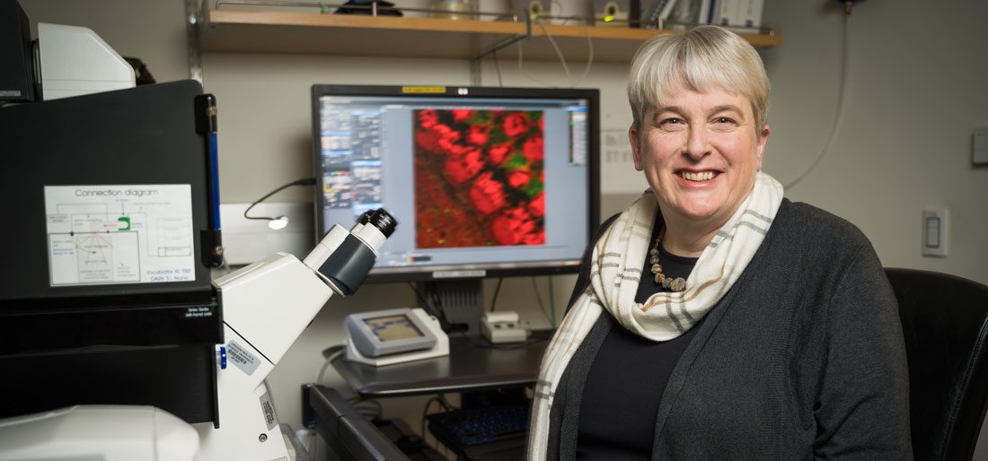
OHSU scientist Stefanie Kaech Petrie, PhD, has been studying cell biology for close to 30 years, but she has not lost her sense of wonder when it comes to microscopes. “Humans are visual creatures. When we see, we believe. When I learned how to use a fluorescence microscope, it transformed the way I understood the world,” said Kaech Petrie.
Kaech Petrie is the director of the Advanced Light Microscopy Core at OHSU’s Jungers Center for Neurosciences Research as well as an associate professor of neurology. She helps researchers from across campus use powerful microscopes to shed light on the structure and function of cells – leading to advancements in treatment for diseases ranging from Alzheimer’s to cancer.
As a child growing up in Switzerland during the 1960s, Kaech Petrie was eager to learn how things worked. Born before the advent of the personal computer, she had one of the first digital cameras and used it constantly. She became increasingly interested in figuring out ways to use computer technology to speed scientific discovery. Her father was a chemist, and she briefly considered following his path.
“I visited the University of Basel for a recruitment program, and the only woman I encountered all day was the cleaning lady. In contrast, in the cell biology department, about one third of the scientists and staff were women. That made my choice,” she said.
Kaech Petrie earned her undergraduate degree and Ph.D. in cell biology from the University of Basel in the 1980s and early 1990s during a time of dramatic growth in the field. Tools capable of looking inside cells started to become available during her training years, and that fascinated her. She went on to specialize in studying brain cells.
“Soon we weren’t just looking inside cells; we were able to do time-lapse imaging, and watch how cells changed over time. The digital revolution in photography made it all possible,” said Kaech Petrie. She was determined to master the technology.
“Soon we weren’t just looking inside cells; we were able to do time-lapse imaging, and watch how cells changed over time. The digital revolution in photography made it all possible.”
Stefanie Kaech Petrie, PhD
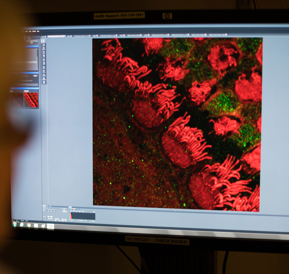
In Andrew Matus’s lab at the Friedrich Miescher Institute in Switzerland, Kaech Petrie studied how neurons (a specific type of brain cell) change shape over time. Neurons are highly specialized, and their shape determines which kinds of cells they talk to.
Neurons can span from the spinal cord to the toe, yet their central bodies and processes are microscopic in size. Subtle changes in their shape are understood to change their function and accompany activities such as memory formation and learning.
To study these changes, Kaech Petrie heavily relied on watching neurons cultured on thin pieces of glass called coverslips. This technique was pioneered by Gary Banker, PhD, a now-retired professor at OHSU who eventually became a Kaech Petrie’s mentor.
When Kaech Petrie was reaching the end of her studies in Switzerland, Banker, who is now retired, invited her to come out to Portland in 2000. She joined his research enterprise to help him answer the question, “How does stuff get shipped around in a neuron?” Together they used light microscopes and time-lapse imaging to help illuminate how neurons transport proteins and other cell components throughout their long extensions.
In 2008, Banker and Kaech Petrie became the first investigators at the newly minted Jungers Center for Neurosciences Research. “With the move to the Jungers Center I was allowed to share my enthusiasm about the transformative nature of modern microscopy with the next generation of neuroscientists,” she said.
“It’s amazing what we built out of that initial seed money from Frank Jungers. I started with one instrument in 2008, and now I have thirteen. We started small, qualified for a federal grant, and now we have a service that is accessible to everyone at OHSU.”
Stefanie Kaech Petrie, PhD
At the time, having a full-time expert focused on lab technology was rare, a luxury that only top research labs could afford. Thanks to philanthropic support from Frank Jungers (after whom the Center was eventually named), OHSU’s neuroscientists could now take advantage of Kaech Petrie’s unique expertise. Funds from private philanthropists and foundations such as Lorry Lokey, the M.J. Murdock Charitable Trust, Truman Collins and the Collins Foundation have also made it possible to support Kaech Petrie’s ambition to build a first-class light microscopy shared resource at OHSU.
“It’s amazing what we built out of that initial seed money from Frank Jungers. I started with one instrument in 2008, and now I have thirteen. We started small, qualified for a federal grant, and now we have a service that is accessible to everyone at OHSU,” said Kaech Petrie.
Kaech Petrie loves her job. It feeds her intense curiosity and allows her to make a significant contribution to science.
“Interacting with so many different scientists – that’s the fun part of my job. I have to understand a little bit about their research, so I am always learning as well as teaching.”

Discovery

Discovery
Discovery
